UV/Vis/NIR for Biomedical Applications
Due to the rapid and accurate UV/Vis spectroscopic capabilities it offers, IDS can be a valuable biomedical tool in the analysis of bacterial and antimicrobial activity in a variety of food, drinks, and biological fluids for medical and healthcare applications. For example, in searching for specific antibiotics, thousands of different bacteria, fungi, and algae must be investigated and their activity analyzed as quickly as possible. This is crucial in order to help prevent various microorganisms from becoming resistant to available antibiotics.[i] IDS can also be a powerful analytical tool for studying the characteristics and population count of bacteria in various liquids. For example, by combining UV/Vis data with Mie scattering theory and Beer-Lambert law, it is possible to determine nucleic and protein content of E. coli cells in water with relative error around 1% compared to traditional plate counting methods.[ii]
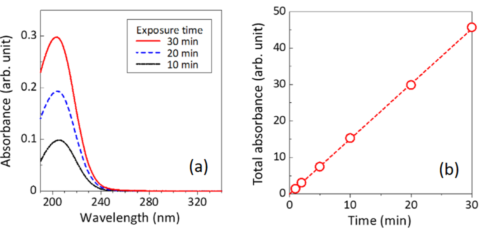
IDS can also be a powerful tool in evaluating the antibacterial properties of liquids, such as plasma-activated water (PAW). UV/Vis techniques have been successfully used in various biomedical applications for analyzing and quantifying the concentrations of reactive oxygen and nitrogen species present in water subjected to different plasma exposure time.iii
Another potential biomedical application of the instrument is in the study of vaccine instability and degradation processes as a function of temperature or storage time. Spectroscopy can be highly effective in monitoring a variety of physical, chemical, and biological pathways that lead to efficacy degradation in vaccine formulations.

UV/Vis absorption spectra of plasma-activated water prepared using different plasma exposure times.[iii]
[i] Slavica B. Ilić, Sandra S. Konstantinović, Zoran B. Todorović. “UV/VIS Analysis and Antimicrobial Activity of Streptomyces Isolates”, Medicine and Biology Vol.12, No 1, 2005, pp. 44 – 46
[ii] Y. Hu et al, “Analytic Method on Characteristic Parameters of Bacteria in Water by Multiwavelength Transmission Spectroscopy”, Journal of Spectroscopy, Volume 2017, Article ID 4039048
[iii] Jun-Seok Oh et al 2018 Jpn. J. Appl. Phys.57 0102B9